Home

Laparoscopic Hysteroscopic Resection

A uterus-preserveing surgery that can directly view the lesion using a fine single-port incision

Laparoscopic Hysteroscopic Resection
Single-port laparoscopy via the umbilicus to remove the fibroid without a scar

Single-port laparoscopy

Fibroid resection performed after entering through a single small opening at the umbilicus


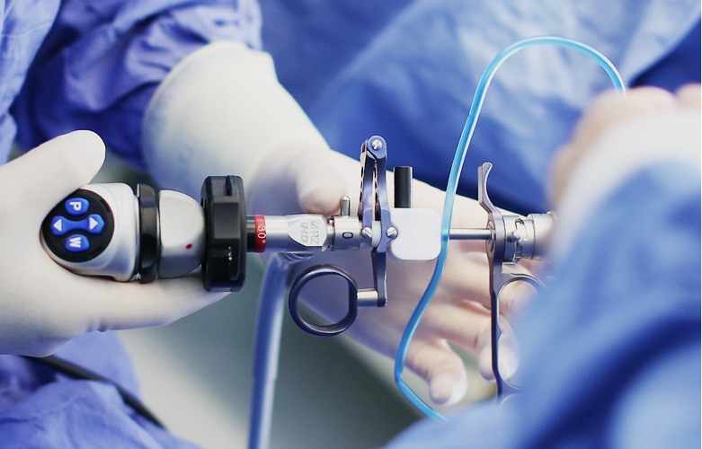
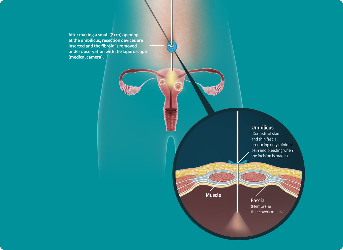
The umbilicus is a vestigial organ where the umbilical cord was once connected. It consists of 'skin + thin fascia' without any abdominal wall muscle or fat layer.
Therefore, an incision leaves little pain and minimal bleeding in this area.Other abdominal areas accompany more severe pain and bleeding when an incision is made as they consist of 'skin + subcutaneous fat + muscle layer'. Since the umbilicus is a wrinkled are with less tension, there is minimal discomfort in the surgery area and its surrounding, and recovery is faster


[ Abdomen image after Single-port laparoscopy ]

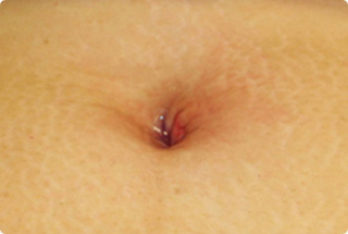
  • Immediately after surgery
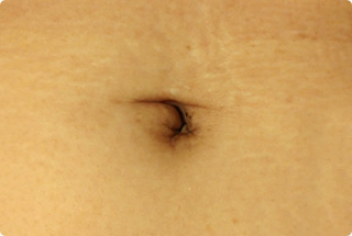
  • 3 months after surgery
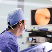
  • Precise surgery
    performed with direct view.

    The laparoscopic camera allows direct view of the tumor, allowing more precise removal of the lesion. State-of-the-art devices provide high resolution images, enabling more delicate surgery. It can treat most of the gynecologic diseases other than fibroids including ovarian tumor and endometriosis.
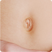
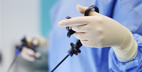
  • 2cm microscopic
    one-point incision in umbilicus

    A single hole is made in the umbilicus to allow entry to the uterus and removal of the fibroid. It is an advanced technique compared to the standard 3~4 hole laparoscopic technique,leaving no hint of surgery as the umbilicus hides the scar.

Features of single-port laparoscopy

Experience and expertise of the surgeon is
crucial due to the higher level of difficulty compared to open surgery


  • Minimal scar and pain

    Scar and pain is minimal as the surgery makes a minimal incision in the umbilicus.Even if it leaves a small surgery mark, it can be hidden by the umbilicus, so you don’t have to be anxious about scar.Also, the hospitalization period is short, 2~3 days in average, allowing fast return to daily life.

  • Little side-effect compared to open surgery

    Minimal invasion, unlike open surgery, leaves little damage to the uterine tissue resulting in a relatively low rate of side effects including bleeding, infection, inflammation and adhesion.

  • Uterine preservation· Immediate treatment effect

    Unlike non-surgical treatments that relieve symptoms gradually, by removing the fibroid tissue itself the effects of surgery are immediately manifested. The uterus is preserved while removing the root cause of symptoms.

Surgery comparison

Open surgery Standard laparoscopy Single-port laparoscopy
Incision Long incision of 10~17cm 3~4 1 cm holes in and around umbilicus 1 2cm hole in umbilicus
Pain ★★★ ★★
Hospitalization 7 days in average 3~5 days in average 2~3 days in average
Return to daily life 6~8 weeks 5~10 days. 5~10 days.

Treatment process

Surgical process

  • Anesthesia and injection of carbon dioxide(CO2) gas

    General anesthesia > CO2gas injection into abdominal cavity with special cannula (To create space for surgery and secure field of vision)

  • Umbilical incision and surgical equipment insertion

    2cm incision in umbilicus > entry point (port) for laparoscopy with high-resolution camera and insertion of 2 surgery devices

  • Removal and suture

    Resection of fibroid while viewing magnified image through monitor > removal of device and gas > suture

After treatment

  • +

    2-year guarantee

    Uterus check-up at 1 week/ 3 months / 6 months /1 year / 2 year (sonography or MRI)

  • +

    Consistent follow-up by doctor

    Your doctor will be in charge of you consultation, tests, and treatment, and be responsible for your lifetime uterine health.

  • +

    Special uterus management program

    We run a management program in collaboration with the Gynecology Center to prevent and treat various uterine diseased other than fibroids

Treatment candidates

Only performed when surgery is necessary in patients unsuitable for non-surgical treatments.

  • Indication
    • Those with symptoms from uterine fibroid (bleeding, anemia, pain, etc.)
    • Patients who are not suitable for MR-HIFU or uterine fibroid embolization and require immediate fibroid resection
    • Pedunculated fibroid (fibroid protruding outward from the uterus connected by a stalk)
    • Broad ligament fibroid(fibroid protruding toward the pelvic ligament), when clinical differential from a malignant tumor of the ovary or adnexa is difficult.
    • History of gynecologic diseases such as ovarian tumor, intrapelvic adhesion, endometriosis, gynecologic tumor, ovarian cyst, ectopic pregnancy
  • Contraindication
    • Fibroid that is rooted deep inside the myometrium
    • Fibroid that cannot be removed easily due to concomitant adenomyosis
    • Excessively large uterine fibroid and adenomyosis

Hysteroscopic(uterine endoscopy) resection

Non-incisional surgery with endoscopy through cervix(vagina)


  • Non-incisional surgery

    It removes the fibroid by entering into the cervix(vagina), without making an incision in the abdomen. Simultaneous treatment and biopsy is offered without damaging the endometrium.
  • Direct removal via endoscope

    After making close observation of the lesion enhanced with hysteroscope, small uterine myoma in cervix and endometrial polyp are removed.
    It is the same principle as removing polyp with colonoscopy.


Features of hysteroscopy

Easy outpatient resection without admission


  • Same-day discharge after resection

    Since no incision is required it does not leave a scar, and bleeding and endometrial damage is minimal.
    It can be performed on an outpatient basis, leaving the hospital on the day of surgery.

  • Applicable to only a limited range of fibroids

    Only fibroids in the uterine cavity can be treated with hysteroscopy.

  • Little complication

    You can experience mild pain or stomach ache. and rare case of infection and vaginal bleeding.
    Infection and vaginal bleeding is a rare complication. You may experience nausea or vomiting as a temporary side effect of anesthesia.

Treatment process

Surgery process

  • Biopsy takes about 5 minutes and whole surgery takes about 30 minutes~ 2 hours depending on the size of uterine fibroid.

  • Anesthesia
    and hysteroscope insertion

    General anesthesia or simple local anesthesia > 3~5mm-thick hysteroscope inserted into uterine cavity via vagina and cervix

  • Resection

    Resection of fibroid while viewing magnified image of uterine cavity through monitor, ablation(burn) or resection if biopsy or resection is necessary > removal of hysteroscope

After treatment

  • +

    2-year guarantee

    Uterus check-up at 1 week/ 3 months / 6 months / 1 year / 2 year (sonography or MRI)

  • +

    Consistent follow-up by doctor

    Your doctor will be in charge of you consultation, tests, and treatment, and be responsible for your lifetime uterine health.

  • +

    Special uterus management program

    We run a management program in collaboration with the Gynecology Center to prevent and treat various uterine diseased other than fibroids

Treatment candidates

Surgery is only performed for patients who are not eligible for non-surgical treatment

  • Indication
    • Fibroid at cervi
    • Submucosal fibroid with small size and endometrial invasion less than 50%
    • Endometrial polyp
  • Contraindication
    • Fibroid located too deep inside myometrium
    • Fibroidthat is not removed easily due to accompanying adenomyosis
    • Excessively large uterine myoma and adenomyosis

Opening Hours

  • Weekdays
    AM 8:30 - PM 5:30
  • Saturday
    AM 9:00 - PM 1:00
    service without lunch time
  • lunch time
    PM 12:30 - PM 1:30

Closed on Sunday & National Holidays.
Opening hours can vary for each medical staff, so please confirm before visiting our hospital

Prev Next